Publications
Evaluating the chemical composition and antitumor activity of Origanum vulgare ssp. hirtum essential l in a preclinical colon cancer model
Georgios Aindelis, Katerina Spyridopoulou, Sotiris Kyriakou, Angeliki Tiptiri-Kourpeti, Mihalis I. Panayiotidis, Aglaia Pappa and Katerina Chlichlia
Int J Mol Sci 2025, 26(10), 4737; https://doi.org/10.3390/ijms26104737
Origanum vulgare ssp. hirtum is an aromatic plant native to various Mediterranean regions and has been traditionally used in folk medicine. This study investigates the chemical composition and the potential antitumor activity of its essential oil in a preclinical model of CT26 colorectal cancer in BALB/c mice. Mice received prophylactic oral administration of the essential oil, and tumor progression, immune modulation, and apoptosis were evaluated. Even treatment with low doses (350 parts per million, ppm in 100 μL final volume) of the essential oil significantly suppressed tumor growth by approximately 44%. This effect correlated with the enhanced expression of antitumorigenic cytokines, including a 2.7-fold increase in type I interferons (IFN), IFN-γ (from 46.5 to 111.9 pg/μL per mg of protein) and tumor necrosis factor alpha (TNF-α) (from 34.5 to 103 pg/μL per mg of protein). Furthermore, the production of granzyme B, a key mediator of cytotoxic immune cell function, was notably increased from 96.1 to 319.6 pg/μL per mg of protein. An elevated activation of caspase 3, a central effector caspase of all apoptotic cascades, was also observed in tumors from oregano-treated mice. These findings suggest that O. vulgare ssp. hirtum essential oil exhibits promising antitumor properties through immune modulation and immunity-mediated apoptosis induction, supporting its potential development as a bioactive compound for cancer prevention or therapy.
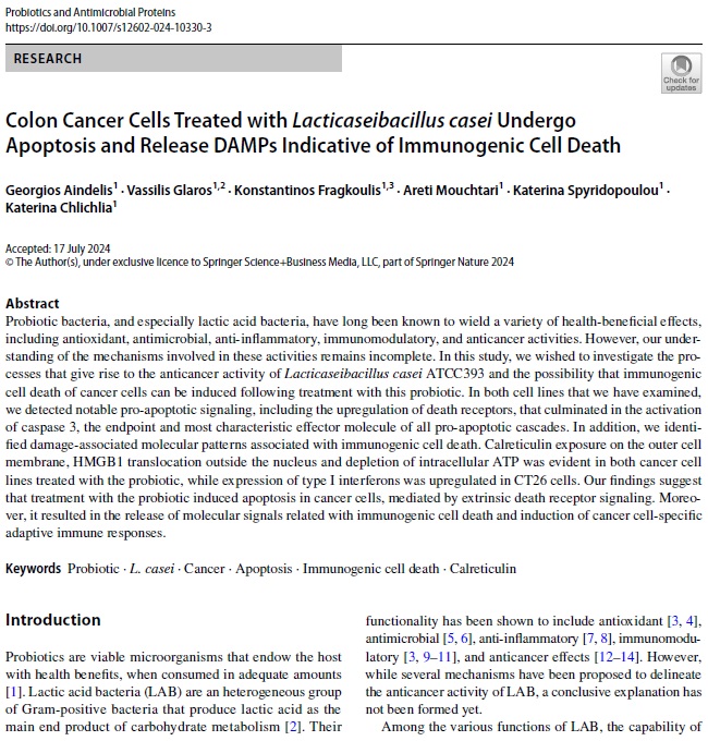
Colon cancer cells treated with Lacticaseibacillus casei undergo apoptosis and release DAMPs indicative of immunogenic cell death
Georgios Aindelis, Vassilis Glaros, Konstantinos Fragkoulis, Areti Mouchtari, Katerina Spyridopoulou and Katerina Chlichlia
Probiotics Antimicrob Proteins 2024, online ahead of print; https://doi.org/10.1007/s12602-024-10330-3
Probiotic bacteria, and especially lactic acid bacteria, have long been known to wield a variety of health-beneficial effects, including antioxidant, antimicrobial, anti-inflammatory, immunomodulatory, and anticancer activities. However, our understanding of the mechanisms involved in these activities remains incomplete. In this study, we wished to investigate the processes that give rise to the anticancer activity of Lacticaseibacillus casei ATCC393 and the possibility that immunogenic cell death of cancer cells can be induced following treatment with this probiotic. In both cell lines that we have examined, we detected notable pro-apoptotic signaling, including the upregulation of death receptors, that culminated in the activation of caspase 3, the endpoint and most characteristic effector molecule of all pro-apoptotic cascades. In addition, we identified damage-associated molecular patterns associated with immunogenic cell death. Calreticulin exposure on the outer cell membrane, HMGB1 translocation outside the nucleus and depletion of intracellular ATP was evident in both cancer cell lines treated with the probiotic, while expression of type I interferons was upregulated in CT26 cells. Our findings suggest that treatment with the probiotic induced apoptosis in cancer cells, mediated by extrinsic death receptor signaling. Moreover, it resulted in the release of molecular signals related with immunogenic cell death and induction of cancer cell-specific adaptive immune responses.
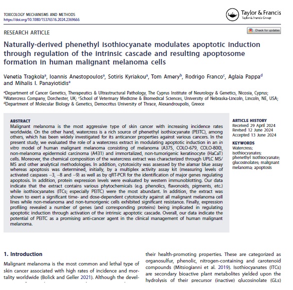
Naturally-derived phenthyl isothiocyanate modulates apoptotic induction through regulation of the intrinsic cascade and resulting apoptosome formation in human malignant melanoma cells
Venetia Tragkola, Ioannis Anetopoulos, Sotiris Kyriakou, Tom Amery, Rodrigo Franco, Aglaia Pappa and Mihalis I. Panayiotidis
Toxicol Mech Methods 2024, 34(9), 985-999; https://doi.org/10.1080/15376516.2024.2369666
Malignant melanoma is the most aggressive type of skin cancer with increasing incidence rates worldwide. On the other hand, watercress is a rich source of phenethyl isothiocyanate (PEITC), among others, which has been widely investigated for its anticancer properties against various cancers. In the present study, we evaluated the role of a watercress extract in modulating apoptotic induction in an in vitro model of human malignant melanoma consisting of melanoma (A375, COLO-679, COLO-800), non-melanoma epidermoid carcinoma (A431) and immortalized, non-tumorigenic keratinocyte (HaCaT) cells. Moreover, the chemical composition of the watercress extract was characterized through UPLC MS/MS and other analytical methodologies. In addition, cytotoxicity was assessed by the alamar blue assay whereas apoptosis was determined, initially, by a multiplex activity assay kit (measuring levels of activated caspases -3, -8 and -9) as well as by qRT-PCR for the identification of major genes regulating apoptosis. In addition, protein expression levels were evaluated by western immunoblotting. Our data indicate that the extract contains various phytochemicals (e.g. phenolics, flavonoids, pigments, etc.) while isothiocyanates (ITCs; especially PEITC) were the most abundant. In addition, the extract was shown to exert a significant time- and dose-dependent cytotoxicity against all malignant melanoma cell lines while non-melanoma and non-tumorigenic cells exhibited significant resistance. Finally, expression profiling revealed a number of genes (and corresponding proteins) being implicated in regulating apoptotic induction through activation of the intrinsic apoptotic cascade. Overall, our data indicate the potential of PEITC as a promising anti-cancer agent in the clinical management of human malignant melanoma.
Isothiocyanates potentiate tazemostat-induced apoptosis by modulating the expression of apoptotic genes, members of polycomb repressive complex 2, and levels of tri-methylating lysine 27 at histone 3 in human malignant melanoma cells
Ioannis Anestopoulos, Ioannis Paraskevaidis, Sotiris Kyriakou, Lambrini E. Glova, Dimitrios T. Trafalis, Sotiris Botaitis, Rodrigo Franco, Aglaia Pappa and Mihalis I. Panayiotidis
Int J Mol Sci 2024, 25(5), 2745; https://doi.org/10.3390/ijms25052745
In this study, we utilized an in vitro model consisting of human malignant melanoma as well as non-tumorigenic immortalized keratinocyte cells with the aim of characterizing the therapeutic effectiveness of the clinical epigenetic drug Tazemetostat alone or in combination with various isothiocyanates. In doing so, we assessed markers of cell viability, apoptotic induction, and expression levels of key proteins capable of mediating the therapeutic response. Our data indicated, for the first time, that Tazemetostat caused a significant decrease in viability levels of malignant melanoma cells in a dose- and time-dependent manner via the induction of apoptosis, while non-malignant keratinocytes were more resistant. Moreover, combinatorial treatment protocols caused a further decrease in cell viability, together with higher apoptotic rates. In addition, a significant reduction in the Polycomb Repressive Complex 2 (PRC2) members [e.g., Enhancer of Zeste Homologue 2 (EZH2), Embryonic Ectoderm Development (EED), and suppressor of zeste 12 (SUZ12)] and tri-methylating lysine 27 at Histone 3 (H3K27me3) protein expression levels was observed, at least partially, under specific combinatorial exposure conditions. Reactivation of major apoptotic gene targets was determined at much higher levels in combinatorial treatment protocols than Tazemetostat alone, known to be involved in the induction of intrinsic and extrinsic apoptosis. Overall, we developed an optimized experimental therapeutic platform aiming to ensure the therapeutic effectiveness of Tazemetostat in malignant melanoma while at the same time minimizing toxicity against neighboring non-tumorigenic keratinocyte cells.
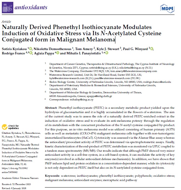
Naturally derived phenethy isothiocyanate modulates induction of oxidative stress via its N-acetylated cysteine conjugated form in malignant melanoma
Sotiris Kyriakou, Nikoletta Demosthenous, Tom Amery, Kyle J. Stewart, Paul G. Winyard, Rodrigo Franco, Aglaia Pappa and Mihalis I. Panayiotidis
Antioxidants 2024, 13(1), 82; https://doi.org/10.3390/antiox13010082
Phenethyl isothiocyanate (PEITC) is a secondary metabolic product yielded upon the hydrolysis of gluconasturtiin and it is highly accumulated in the flowers of watercress. The aim of the current study was to assess the role of a naturally derived PEITC-enriched extract in the induction of oxidative stress and to evaluate its anti-melanoma potency through the regulation of its metabolism with the concurrent production of the N-acetyl cysteine conjugated by-product. For this purpose, an in vitro melanoma model was utilized consisting of human primary (A375) cells as well as metastatic (COLO-679) malignant melanoma cells together with non-tumorigenic immortalized keratinocytes (HaCaT). Cytotoxicity was assessed via the Alamar Blue assay whereas the antioxidant/prooxidant activity of PEITC was determined via spectrophotometric assays. Finally, kinetic characterization of the end-product of PEITC metabolism was monitored via UPLC coupled to a tandem mass spectrometry (MS/MS). Our results indicate that although PhEF showed very minor antioxidant activity in a cell-free system, in a cell-based system, it can modulate the activity of key enzyme(s) involved in cellular antioxidant defense mechanism(s). In addition, we have shown that PhEF induces lipid and protein oxidation in a concentration-dependent manner, while its cytotoxicity is not only dependent on PEITC itself but also on its N-acetylated cysteine conjugated form.
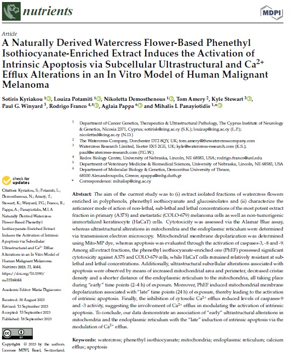
A naturally derived watercress flower-based phenethyl isothiocyanate-enriched extract induces the activation of intrinsic apoptosis via subcellular ultratructural and Ca2+ efflux elterations in an in vitro model of human malignant melanoma
Sotiris Kyriakou, Louiza Potamiti, Nikoletta Demosthenous, Tom Amery, Kyle Stewart, Paul G. Winyard, Rodrigo Franco, Aglaia Pappa and Mihalis I. Panayiotidis
Nutrients 2023, 15(18), 4044; https://doi.org/10.3390/nu15184044
The aim of the current study was to (i) extract isolated fractions of watercress flowers enriched in polyphenols, phenethyl isothiocyanate and glucosinolates and (ii) characterize the anticancer mode of action of non-lethal, sub-lethal and lethal concentrations of the most potent extract fraction in primary (A375) and metastatic (COLO-679) melanoma cells as well as non-tumorigenic immortalized keratinocyte (HaCaT) cells. Cytotoxicity was assessed via the Alamar Blue assay, whereas ultrastructural alterations in mitochondria and the endoplasmic reticulum were determined via transmission electron microscopy. Mitochondrial membrane depolarization was determined using Mito-MP dye, whereas apoptosis was evaluated through the activation of caspases-3, -8 and -9. Among all extract fractions, the phenethyl isothiocyanate-enriched one (PhEF) possessed significant cytotoxicity against A375 and COLO-679 cells, while HaCaT cells remained relatively resistant at sub-lethal and lethal concentrations. Additionally, ultrastructural subcellular alterations associated with apoptosis were observed by means of increased mitochondrial area and perimeter, decreased cristae density and a shorter distance of the endoplasmic reticulum to the mitochondria, all taking place during “early” time points (2–4 h) of exposure. Moreover, PhEF induced mitochondrial membrane depolarization associated with “late” time points (24 h) of exposure, thereby leading to the activation of intrinsic apoptosis. Finally, the inhibition of cytosolic Ca2+ efflux reduced levels of caspases-9 and -3 activity, suggesting the involvement of Ca2+ efflux in modulating the activation of intrinsic apoptosis. To conclude, our data demonstrate an association of “early” ultrastructural alterations in mitochondria and the endoplasmic reticulum with the “late” induction of intrinsic apoptosis via the modulation of Ca2+ efflux.
Chemical characterization and biological evaluation of Epilobium parvifloum extracts in an in vitro model of human malignant melanoma
Sotiris Kyriakou, Venetia Tragkola, Ioannis Paraskevaidis, Mihalis Plioukas, Dimitrios T. Trafalis, Rodrigo Franco, Aglaia Pappa and Mihalis I. Panayiotidis
Plants 2023, 12(8), 1590; https://doi.org/10.3390/plants12081590
Malignant melanoma is an aggressive type of skin cancer characterised by high metastatic capacity and mortality rate. On the other hand, Epilobium parviflorum is known for its medicinal properties, including its anticancer potency. In this context, we aimed to (i) isolate various extracts of E. parviflorum, (ii) characterize their phytochemical content, and (iii) determine their cytotoxic potential in an in vitro model of human malignant melanoma. To these ends, we utilized various spectrophotometric and chromatographic (UPLC-MS/MS) approaches to document the higher content of the methanolic extract in polyphenols, soluble sugars, proteins, condensed tannins, and chlorophylls-a and -b as opposed to those of dichloromethane and petroleum. In addition, the cytotoxicity profiling of all extracts was assessed through a colorimetric-based Alamar Blue assay in human malignant melanoma (A375 and COLO-679) as well as non-tumorigenic immortalized keratinocyte (HaCaT) cells. Overall, the methanolic extract was shown to exert significant cytotoxicity, in a time and concentration-dependent manner, as opposed to the other extracts. The observed cytotoxicity was confined only to human malignant melanoma cells, whereas non-tumorigenic keratinocyte cells remained relatively unaffected. Finally, the expression levels of various apoptotic genes were assessed by qRT-PCR, indicating the activation of both intrinsic and extrinsic apoptotic cascades.
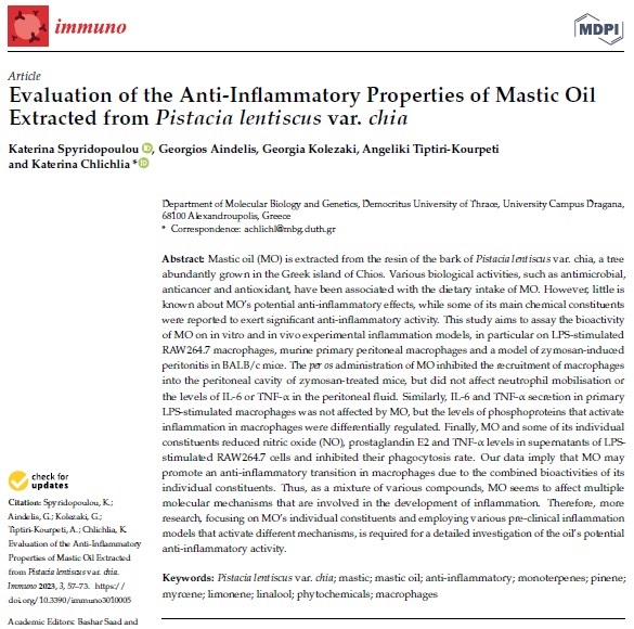
Evaluation of the anti-inflammatory properties of mastic oil Extracted from Pistacia lentiscus var. chia
Katerina Spyridopoulou, Georgios Aindelis, Kolezaki Georgia, Angeliki Tiptiri-Kourpeti and Katerina Chlichlia
Immuno 2023, 3(1), 57-73; https://doi.org/10.3390/immuno3010005
Mastic oil (MO) is extracted from the resin of the bark of Pistacia lentiscus var. chia, a tree abundantly grown in the Greek island of Chios. Various biological activities, such as antimicrobial, anticancer and antioxidant, have been associated with the dietary intake of MO. However, little is known about MO’s potential anti-inflammatory effects, while some of its main chemical constituents were reported to exert significant anti-inflammatory activity. This study aims to assay the bioactivity of MO on in vitro and in vivo experimental inflammation models, in particular on LPS-stimulated RAW264.7 macrophages, murine primary peritoneal macrophages and a model of zymosan-induced peritonitis in BALB/c mice. The per os administration of MO inhibited the recruitment of macrophages into the peritoneal cavity of zymosan-treated mice, but did not affect neutrophil mobilisation or the levels of IL-6 or TNF-α in the peritoneal fluid. Similarly, IL-6 and TNF-α secretion in primary LPS-stimulated macrophages was not affected by MO, but the levels of phosphoproteins that activate inflammation in macrophages were differentially regulated. Finally, MO and some of its individual constituents reduced nitric oxide (NO), prostaglandin E2 and TNF-α levels in supernatants of LPS-stimulated RAW264.7 cells and inhibited their phagocytosis rate. Our data imply that MO may promote an anti-inflammatory transition in macrophages due to the combined bioactivities of its individual constituents. Thus, as a mixture of various compounds, MO seems to affect multiple molecular mechanisms that are involved in the development of inflammation. Therefore, more research, focusing on MO’s individual constituents and employing various pre-clinical inflammation models that activate different mechanisms, is required for a detailed investigation of the oil’s potential anti-inflammatory activity.
Conferences
Backround by pikisuperstar on Freepik
Contact bioactivescreen.duth@gmail-com
The project was supported by the Hellenic Foundation for Research and Innovation (H.F.R.I.) under the “1st Call for H.F.R.I. Research Projects to support Faculty members and Researchers and the procurement of high-cost research equipment” (Project Number: HFRI-FM17C3-2007)


